
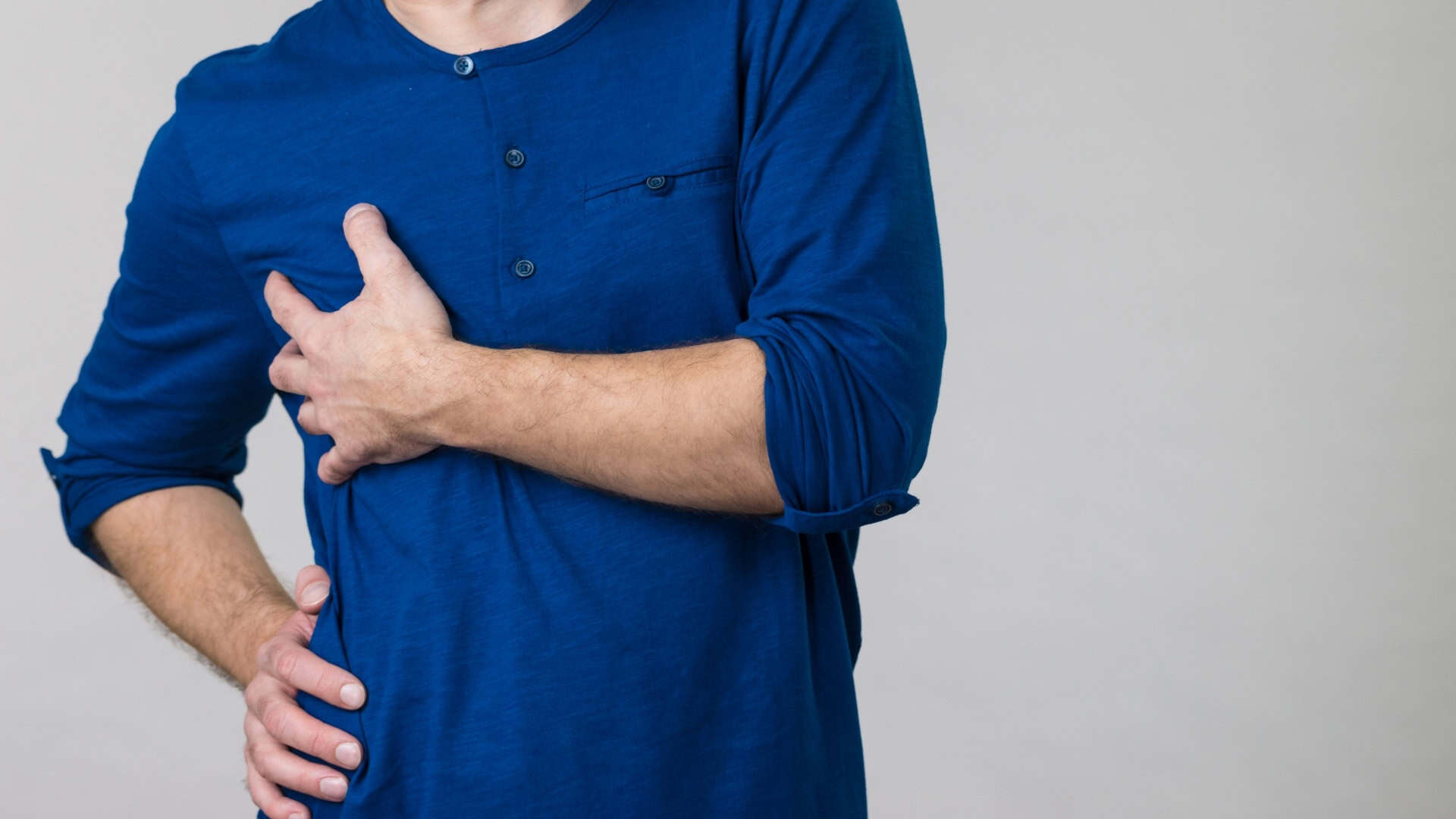
The Rib Condition That Gets Misdiagnosed for Years
You might assume rib pain that lingers for months or even years is just muscle strain or anxiety, especially when every test comes back “normal.” Yet certain chest-wall conditions, like Tietze syndrome, hide in plain sight and get mistaken for everything from heart trouble to simple costochondritis. If your pain is sharp, tender to the touch, and still doesn’t make sense, there’s a good chance something important is being missed…
Tietze Syndrome: What It Is and Why It’s Missed
Tietze syndrome is an uncommon chest wall condition in which one or more costal cartilages most frequently at the 2nd or 3rd ribs become inflamed. This inflammation produces localized, sharp pain and a clearly palpable, tender swelling where the rib meets the breastbone (sternum). The affected area may feel warm and can appear slightly discolored.
Pain typically worsens with movements that stress the chest wall, such as coughing, sneezing, deep breathing, twisting, or lifting.
The condition is frequently overlooked or misclassified because chest pain often prompts an initial evaluation for cardiac or pulmonary causes. When investigations such as ECG, blood tests, and imaging don't indicate heart or lung disease, the pain is commonly attributed to nonspecific “costochondritis,” even when focal swelling is present.
In contrast to typical costochondritis, Tietze syndrome is characterized by visible or palpable swelling at the involved costochondral junction.
Diagnosis is primarily clinical and is based on the characteristic pattern of pain and localized swelling, along with the exclusion of other serious causes of chest pain.
Imaging studies such as ultrasound or MRI may help confirm inflammation and rule out alternative diagnoses, but they aren't always required.
Symptom improvement with rest and anti-inflammatory treatment can further support the diagnosis, although response to therapy isn't specific to Tietze syndrome.
Tietze vs Costochondritis vs Slipping Rib: Key Differences
While all three conditions can cause persistent chest or rib pain, they involve different structures and locations in the rib cage.
Tietze syndrome typically causes a single, localized, tender swelling where an upper rib (often the second or third) meets the breastbone (sternum). The area may feel warm or visibly raised, and pain is usually well localized to that swollen joint.
Costochondritis also involves the cartilage where ribs join the sternum, but it usually affects multiple sites and doesn't cause visible swelling. The pain is reproducible when pressing on the affected costochondral junctions, but the overlying skin and tissue generally look normal.
Slipping rib syndrome affects the lower “false” ribs (usually ribs 8–10), where there's excessive movement of the rib tips under the costal arch. This can lead to intermittent sharp pain, sometimes accompanied by a clicking or popping sensation, often triggered by twisting, bending, or deep breathing. Diagnosis may be supported by dynamic ultrasound and the hook maneuver, where a clinician reproduces symptoms by lifting the lower rib margin. Management typically starts with activity modification, physical therapy, and pain control; in more severe or persistent cases, options can include nerve blocks or surgical stabilization.
Of the three, slipping rib syndrome is the one most likely to go unnamed the longest. London thoracic surgeon Mr Marco Scarci, who treats it regularly, points out that patients commonly wait several years and see five or more specialists before anyone performs the hooking maneuver, a bedside test that takes under five minutes. His guide to the condition covers how that examination works, why standard scans keep coming back clear, and where cartilage resection fits once conservative treatment has been exhausted.
Common Symptoms of Misdiagnosed Rib and Chest-Wall Pain
For many people, persistent rib or chest‑wall pain presents as a small, localized area of discomfort often on one side of the front or upper chest that becomes more noticeable with twisting, torso rotation, or pushing movements such as pushups or bench press.
The pain may feel sharp or stabbing with movement, coughing, sneezing, or deep breathing, followed by a more constant, dull ache.
It frequently centers around a single rib or rib‑cartilage junction and may worsen with direct pressure or when holding certain postures.
When the lower ribs are involved, some individuals report popping or clicking sensations.
Stretching the area between the ribs (the intercostal spaces) can be painful, even when X‑rays or other imaging studies don't show structural abnormalities.
What’s Actually Causing Tietze and Slipping Rib Pain?
Although both conditions present as persistent chest wall pain, they result from different mechanical problems involving the ribs and their cartilage.
In Tietze syndrome, the primary issue is localized inflammation and swelling where an upper rib most often the second or third joins the sternum (the costochondral junction). This inflammation can make the area visibly enlarged and tender.
Movements or actions that load or stretch this region, such as twisting the torso, coughing, sneezing, or applying direct pressure, can aggravate the irritated cartilage and nearby nerves, leading to increased pain.
In slipping rib syndrome, the problem typically involves the lower ribs, usually ribs 8–10, which are connected to the cartilage rather than directly to the sternum.
One of these rib tips can become hypermobile or unstable and may partially slip beneath the costal margin (the lower edge of the rib cage). This abnormal movement can mechanically compress or irritate the intercostal tissues and nerves during bending, twisting, deep breathing, or lifting and carrying objects, producing sharp, sometimes sudden pain.
How Doctors Diagnose Rib-Related Chest Pain (When It’s Not Your Heart)
Because chest pain can indicate a heart attack or other medical emergency, clinicians first prioritize ruling out life-threatening cardiac and pulmonary causes. This typically involves assessing vital signs, performing an electrocardiogram (EKG), and examining the heart and lungs.
Additional tests, such as blood work or imaging, may be ordered if indicated.
Once serious conditions are considered unlikely, the evaluation shifts toward the ribs and surrounding structures. The clinician may carefully press along each rib and where the ribs meet the breastbone (sternum) to see whether they can reproduce the exact area of pain.
They may also guide you through specific movements such as twisting, reaching forward, or arching your back to identify motions that trigger or worsen the discomfort.
In some cases, dynamic ultrasound is used while you move, allowing the examiner to observe the ribs, cartilage, and nearby soft tissues in real time for signs of slipping, inflammation, or other abnormalities.
If the source of pain is still unclear, a targeted local anesthetic (numbing) injection around a suspected rib or nerve may be used. Significant pain relief after the injection can help confirm that area as the primary source of symptoms.
Why Your Scans and Tests Can Look “Normal” for Years
Once heart and lung conditions have been ruled out, a common next issue is that standard imaging appears normal even though the pain continues. This occurs because slipping ribs and related conditions are often dynamic problems.
When you're lying still, X‑rays, CT scans, and MRIs may show no clear abnormality, so the source of pain isn't obvious.
However, during movement such as twisting, coughing, or bending the rib tip can shift position and temporarily narrow the space between ribs. Dynamic ultrasound, performed while you move or after a specific provoking maneuver, can sometimes capture this change and identify the problem area.
In addition, targeted clinical tests, such as the hook maneuver or a diagnostic intercostal nerve block, can help confirm which rib is involved, even when previous imaging results have all been reported as normal.
Treatments That Work for Tietze and Slipping Rib Syndrome
When rib pain persists for months or longer, appropriate treatment depends on whether the primary issue is inflammatory (as in Tietze syndrome) or mechanical (as in slipping rib syndrome).
In Tietze syndrome, management usually begins with rest, temporary reduction of aggravating activities, and nonsteroidal anti-inflammatory drugs (NSAIDs) to reduce costochondral joint inflammation and tenderness. In some settings, additional options such as extracorporeal shockwave therapy directed to the affected costochondral region and targeted soft-tissue or mobility techniques may be used to improve local mechanics and relieve symptoms, although evidence for these approaches is more limited.
For slipping rib syndrome, dynamic ultrasound can help visualize abnormal rib motion, and diagnostic intercostal nerve blocks may help confirm which rib segment is responsible for the pain. Initial management commonly includes activity modification, physical therapy, and analgesic medication. If conservative treatment doesn't provide adequate relief and symptoms are clearly linked to rib hypermobility, surgical stabilization (such as fixation or partial rib resection) can be considered and may provide more durable symptom control in selected patients.
Exercising Safely and Managing Triggers With Chronic Rib Pain
Even with chronic rib pain, many people can remain active by identifying and modifying specific symptom triggers. Pay attention to movements that reliably reproduce pain, such as twisting, rounding the upper back, taking deep breaths, coughing or sneezing, or performing exercises that involve heavy pushing (for example, bench press, dips, or pushups).
Reduce, modify, or temporarily stop activities that provoke symptoms, then gradually reintroduce them within pain‑free or minimally symptomatic ranges. Emphasize stretching tight chest‑wall and surrounding soft tissues, strengthening the shoulder and trunk stabilizing muscles, and practicing rib‑cage stabilization exercises, rather than continuing to exercise through sharp, localized pain.
If the pain is focused at the lower rib margins and feels like a small, hard area or “lump,” especially near the rib tips, discuss the possibility of slipping‑rib syndrome with a clinician and ask about appropriate physical examination and dynamic imaging or testing.
When to Push for a Second Opinion on Persistent Rib Cage Pain
Persistent rib pain that interferes with training or daily activities warrants further medical evaluation. If you have been treated for “costochondritis” without meaningful improvement and your symptoms have continued for months or years, it's reasonable to request a second opinion. The NHS overview of costochondritis is a useful benchmark here: if what you are experiencing doesn't match the description, that mismatch is worth raising directly at the appointment.
Provide your clinician with a detailed description of what provokes the pain, such as twisting, bending, lifting, coughing, or taking a deep breath. Note any sensations of clicking, popping, or shifting near the lower rib margins. Ask whether conditions such as slipping rib syndrome or other rib-cage–related disorders could explain your symptoms.
You can also ask about specific bedside assessments, including controlled torso rotation or a “hook” maneuver to evaluate rib mobility, and whether a dynamic (movement-based) ultrasound performed by an experienced clinician might be appropriate. Seeking timely reassessment may help identify under-recognized chest wall conditions and guide more targeted management.
Conclusion
When your rib pain keeps getting brushed off or mislabeled, you’re not being “dramatic” you’re dealing with something real that deserves answers. Now you know how Tietze, costochondritis, and slipping rib differ, why scans can look normal, and what treatments actually help. Use that knowledge to track patterns, protect your triggers, and ask sharper questions. If pain persists or limits your life, push for a second opinion. You’re allowed to insist on real relief.
Awards and
Certifications
